Translation of the original German introduction by Ernst Haeckel:
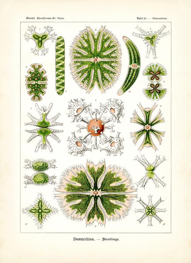
Division of Protophyta (Urpflanzen); - class of Pauosporata (Algarien); - subclass of Desmidiacea (Kosmarien).
Desmidiacea (Kosmarien or Zierdinge) form a varied subclass within the division of unicellular Protophyta, in fact, within the class that knows no ‘flicker movement’ (Algaria). All Desmidiacea dwell in freshwater (mainly in marsh); they are distinguished by the delicate symmetrical shape of their cell membrane or ‘cellulose shell’ that is frequently armed with thorny prickles. The living plasma body (Cytosoma) that dwells in this shell covers a green pigment body (Chromatell) of tender shape; this body usually consists of two symmetrical ‘chlorophyll discs’ with radial lopes (fig. 12 and 13), more rarely of more discs (fig. 10), sometimes of a ‘spiral band’ (fig. 9). Usually several shiny ‘protein crystals’ (Pyrenoide) rest inside the Chromatell. In the centre of each cell lies the plain nucleus. Reproduction of Desmidiacea follows a peculiar dual way: at first through simple cell division and secondly through pairing. In simple cell division (fig. 6, 7) both halves of the symmetrical cell pinch off each other, with each half forming a new cell half at the ‘separation plane’ through ‘supplementation growth’; the new half grows until it reaches size and shape of the old cell. In pairing (conjugation or copulation), however, two cells lie one upon the other (fig. 2, 3 and 4); both flaps of the shell halves of each cell detach from each other and allow the two now free cell bodies (Cytosoma) to coalesce.
The thus created (usually nodular) new cell – Zygospore (Paarling or Jochspore) – surrounds itself with a membrane that is usually armed with radial flagella (fig. 5). Later the cell leaves this cover.
Translation by VR Translators Bangalore
We've scanned the original lithography at 1200dpi on the Epson A3 scanner of A3 scanner huren. You can download a 400dpi JPEG here.