Translation of the original German introduction by Ernst Haeckel:
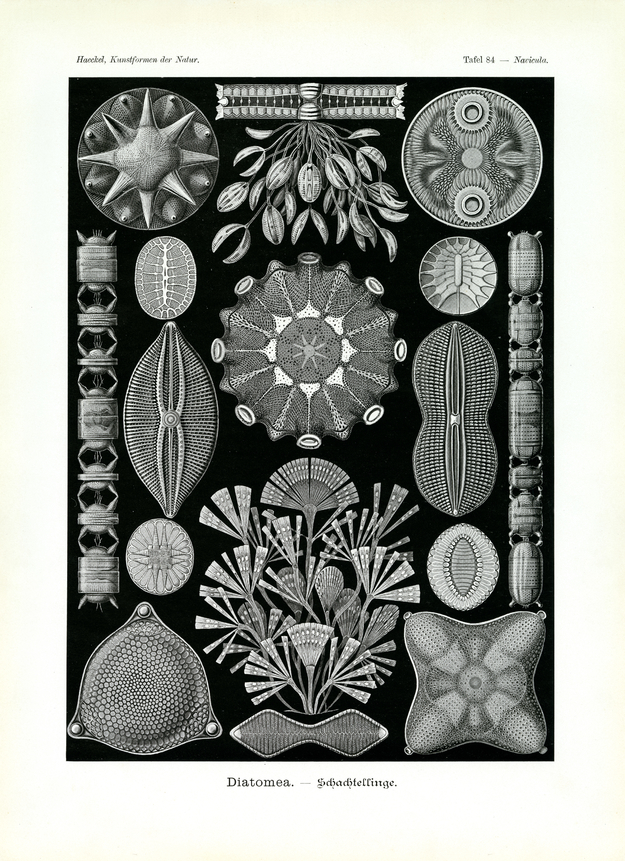
Division of Protophyta (Urpflanzen); main class of Algaria; class of Diatomea or Bacillaria (Schachtel- or Kiesel-Algaria).
The varied species of Diatomea or Schachtellinge illustrated on this plate are, to a large extent, or individually living ‘celled-ones’, just like all species presented on plate 4. In addition, the illustration here presents four distinct species of Coenobia or ‘cell-union’, composed of numerous convivial cells that originate in repeated division of a ‘mother cell’. These “cell-cormidia” or “cell-colonies” are partly free and string-shaped with all convivial cells organized in a row: ‘string-unions’ (or Catenal-Coenobia, fig. 7, 9); partly sessile, bush- or tree-shaped, fixed on branched-off gelatinous shafts: ‘tree-unions’ (or Arboral-Coenobia, fig. 1, 14). The number of particular cell-individuals that live united in such a colony, may amount to several thousands in the case of big Coenobia.
Although the frame of the living Diatomea-cell is most simple (a rounded plasma body containing an individual nucleus in the centre), the shape of the lime shell deposited by it is most diverse, distinguished by an extraordinary delicate and regular sculpture. All these lime shells have a characteristic ‘case-frame’ in common, with both flaps of the lime shell loosely connected like a box and its lid. The upper, slightly bigger half, the case-lid, uses a broad rim, the ‘belt’, to take hold of the rim of the lower, slightly smaller half (of the case-flap). The firm and most particularly shaped lime shell therefore offers two very different perspectives; seen from the main or basal side a usually most differentiated and delicate sculpture is seen (at the horizontal base of the case as well as at the top of the flap) whereas the more narrow lateral side, also known as ‘belt-side’ or ‘belt-band-side’, usually appears more simple.
Innumerable small pores that perforate the lime-shell are most regularly organized into radially distributed planes separated by protruding bars. Many species are two-radiant (fig. 5, 6, 10, 11), others three-radiant (fig. 15), four-radiant (fig. 16), six- or eight-radiant (fig. 8). (Cf. plate 4 with explanation.) At times the distinct ‘threat-drawing’ appears fixed in the delicate sculpture of the lime shell, showing so-called mitosis in the usual, indirect cell-division (fig. 3). The living content of the lime-shell (Protoplast) usually appears yellow or yellowish-brown in living Diatomea-cells due to particular ‘pigment-grains’ (Chromatella) distributed in the ‘plasma-net’. The round nucleus (Nucleus or Karyon) is positioned in the centre of the cell.
Translation by VR Translators Bangalore
We've scanned the original lithography at 1200dpi on the Epson A3 scanner of A3 scanner huren. You can download a 400dpi JPEG here.