Translation of the original German introduction by Ernst Haeckel:
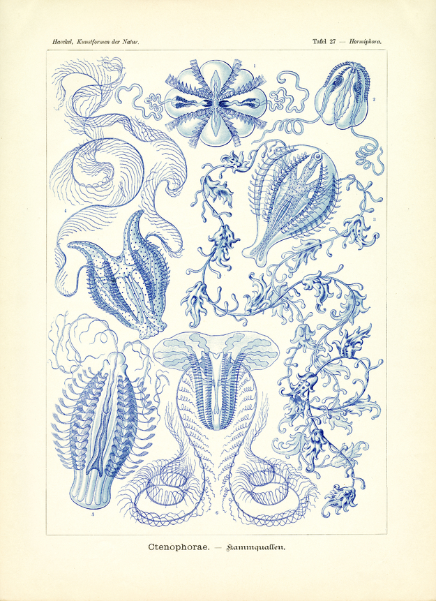
Phylum of Cnidaria (Nesseltiere); - subclass of Ctenophorae (Kammquallen); - legion of Cannocteniae (with simple rip canals); - order of Cydippeae (Saccaten).
Ctenophorae (Kammquallen or Rippenquallen) form a very peculiar subclass within the phylum of Cnidaria; they are probably most closely related to Craspedotae and originate from a branch of Anthomedusae (plate 6, fig. 1-4). All Ctenophorae live swimming in the open sea and are distinguished by extraordinary tenderness of their soft, gelatinous body. The water content of the body usually amounts to 96 to 99 percent, thus, only 1 to 4 percent (or even less) cover the weight of animal tissue. Most often the vitreous body is perfectly transparent allowing the perception of the internal structure without difficulty. The size is most variable; it amounts to a few millimeters only in the smallest species, to more than a meter in the biggest.
The geometric basic form of the body is most peculiar with the outer overall shape being almost nodular or ovoid, at times in the form of a pear or a melon. Usually the inner organs and the outer appendices of the body are structured in such a way that the abstract geometrical basic form is four-jetted and at the same time double-edged (rhombus pyramid, meaning a quadrangular pyramid with a rhombus as base). Of the three distinct axis shafts that sit vertically on one another, thus determining the basic shape, the first one, main axis, is poled unevenly; at the lower, oral pole (‘mouth pole’) the mouth opening is located, at the upper, aboral pole (‘funnel pole’) the funnel and the ‘nerve nodes’ together with the sense organ. The other two axis shafts are homopolar; the laterally compressed pharynx lies in the shorter (sagittal) axis (in fig. 1 seen from above, vertically); in the longer axis, to the right and to the left, two long tentacles or ‘capturing filaments’ are seen that can be retreated into special tentacle pockets (in fig. 1 horizontally).
A most distinctive feature in Ctenophorae is their peculiar locomotion apparatus that gave the subclass its name. It consists of eight adrial ‘cilia combs’ or ‘flickering ribs’ passing in shallow meridian arches from one pole of the vertical main axis to the other. Each comb consists of a row on swinging tender ‘cilia lamellae’ that sit on the broad rim of the main upper plane and are split into numerous delicate cilia. Whenever the sun shines on these slowly swimming animals the interference of light brings about the most magnificent play of colours of a steadily changing rainbow. With the help of the arbitrary movements of these ‘cilia ribs’, that beat as consistently as the line of oars in a galley, the delicate Ctenophorae drift slowly in the sea. The inner body structure is similar to that of Medusae. The flexible mouth opening (at the base) leads into a broad pharynx cavity; it changes at the top into a smaller stomach cavity, the so called funnel. At the top this funnel splits into two funnel canals that embrace the ‘nerve nodes’ located there, the ‘parting brain’ along with the attached nodular sense organ at the ‘parting pole’ (fig. 3 und 4). Two massive food canals leave laterally, splitting twice in the form of a fork; thus each of the eight ‘cilia combs’ receives a ‘rib canal’; out of the wall of the ‘rib canal’ Gonads develop in such a way that in each canal a male gonad is found on one side and a female one on the other.
Translation by VR Translators Bangalore
We've scanned the original lithography at 1200dpi on the Epson A3 scanner of A3 scanner huren. You can download a 400dpi JPEG here.