Translation of the original German introduction by Ernst Haeckel:
Phylum of Cnidaria (Nesseltiere); - subclass of Acraspedae (Lappenquallen); - order of Discomedusae (Scheibenquallen); - suborder of Rhizostomae (Wurzelmündigen).
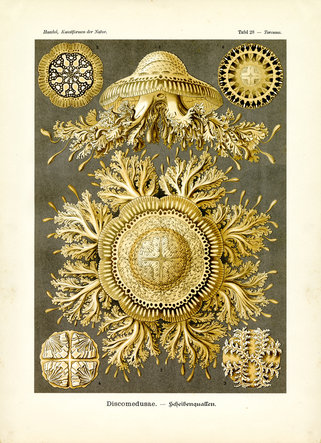
Rhizostomae (Wurzelmündige) form the third and youngest of the three suborders of Discomedusae (Scheibenquallen), distinguished due to the fact that in the adult Medusa the central mouth opening has become fully grown over. During its youth the animal has the original mouth formation of the Cannostomae (Rohrmündigen, plate 18); the mouth is located at the lower end of a mouth tube emanating from the centre of the lower umbrella plane; it is split into four short lopes (plate 18, fig. 2-5).
Later on these four frilled ‘mouth lopes’ develop into four powerful flexible arms, the richly wrinkled ‘mouth arms’ or ‘mouth curtains’ that identify the suborder of Semostomae (Fahnenmündige, plate 8). Rhizostomae originate from these Semostomae by splitting four ‘mouth banners’ in the form of a fork into two branches each, and by growing together of the numerous, juxtaposed folds of the ‘mouth frills’ of these eight strong ‘mouth arms’. Similar tubes come into being when the folds of a strongly stiffened shirt collar or a round collar are glued at the points of contact. Food reaches them (Rhizostomae) through the numerous small openings (‘small sucking mouths’) at the outer end of the tubes, further transported through their inner openings into the central gastrovascular cavity. The central part of the intermediate mouth however becomes completely overgrown; the cross-shaped connation seam of this frizzled ‘mouth cross’ remains intact (fig. 3). Usually, the numerous branches of the eight frizzy, thick ‘mouth arms’ branch off so severely that cauliflower-like formations come into being with thousands of small ‘sucking mouths’. Frequently peculiar piston-shaped bubbles are fixed in between these (fig. 1 and 2).
The hat-like arched or flatly disc-shaped umbrella of the ‘root mouthed’ Discomedusa includes the central gastrovascular cavity in the centre with usually 16 branched ‘jet canals’ towards the rim of the umbrella. Below the stomach cavity lie four crescent-shaped or triangular Gonads, fixed to delicate, wrinkled bands (Gonads, fig. 4). In between them the lower space of the gastrovascular cavity forms a perpendicular cross (fig. 2, 4, 6). The arched outer or upper umbrella plane (Exumbrella) of many of the Rhizostomae is ornated by light coloured (white or yellow) spots regularly distributed and contrasting poignantly against the dark (usually yellow or red, violet or blue) background of the gelatinous umbrella (fig. 1, 2, 5, 6). – The rim of the umbrella is distinguished in Rhizostomae by the fact that the flexible thread-shaped tentacles common in other Medusae have disappeared due to regression. Usually the rim of the umbrella is notched delicately or split into numerous fine lopes. 8 – 16 ‘sensing buds’ of Rhophalia are sitting in between these in deep incisions; each is composed of an eye, a ‘hearing bubble’ and a ‘smelling groove’.
Translation by VR Translators Bangalore
We've scanned the original lithography at 1200dpi on the Epson A3 scanner of A3 scanner huren. You can download a 400dpi JPEG here.